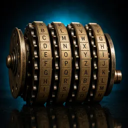
AGENTEX runs a 34-codon genetic code outside a living cell
AGENTEX pairs altered tRNA ends with matching ribosomes in a cell-free system and runs compressed 34-codon codes. A separate eLife study tests AroMIP, which predicts membrane insertion by aromatic motifs from sequence. Manipulated marketing images for commercial antibodies show why validation remains inseparable from laboratory tools: programmability matters only alongside reliable measurement and an honest account of performance.
Science··Night
A second connection path for the genetic code
The standard genetic code maps 64 codons onto 20 canonical amino acids. The peer-reviewed Nature study built AGENTEX to test alternative mappings without first rewriting a living genome. The system pairs synthetic tRNAs carrying altered 3′ ends instead of the usual CCA with ribosomes whose large subunit has been changed, then screens libraries robotically. Most Escherichia coli aminoacyl-tRNA synthetases could charge the altered tRNAs. Using that flexibility, the team ran two compressed codes mapping 34 synthetases onto 34 codons alongside the standard code, added non-standard amino acids and reassigned up to three codons. The result belongs to a cell-free experiment; no living cell carries these codes.[1]
A sequence-level prediction of membrane insertion
The peer-reviewed eLife version of record asks whether the sequence around an aromatic motif in a disordered protein can predict entry into a membrane's oily interior. The researchers first compared 10 nine-residue motifs using all-atom simulations and the PPM method for positioning proteins in membranes. They then scanned 1.2 million sequences centred on phenylalanine, tryptophan or tyrosine and built a mathematical model called AroMIP. Reported test-set accuracies on motifs drawn from the human proteome were 91.2 per cent, 92.0 per cent and 99.7 per cent, respectively. Those figures measure how well the model reproduces labels produced by PPM, rather than directly observed membrane insertion across all human proteins.[2]
Manipulated antibody images push the experiment back onto the lab
The reliability of a laboratory tool also depends on the evidence available to the researcher who buys it. In the sweep reported by Ars Technica, Reese Richardson and Sholto David used partly automated methods to examine stained-cell and western-blot images that antibody suppliers publish as marketing. They found problematic changes associated with about 17,500 antibodies from 16 companies; nearly 7 per cent of the examined images showed signs ranging from erased background to copied regions. When an antibody fails to produce the expected result, buyers often question their own experimental conditions first. An image that overstates performance can therefore turn into weeks of further adjustment, wasted reagents and a false starting assumption.[3]
Related columns
For more information on this topic, you can read the related columns.